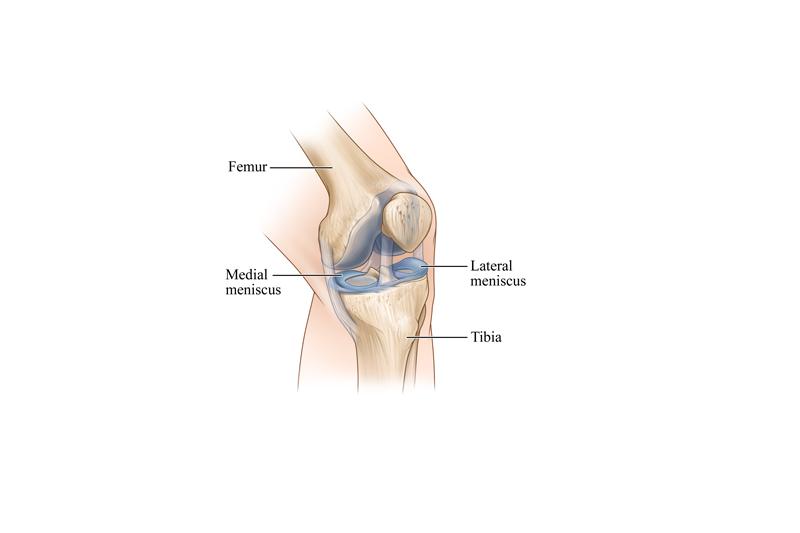
Injury to the knee-joint may produce acute traumatic synovitis or haemoarthritis. The two conditions must be differentiated. In traumatic synovitis, there is synovial effusion due to the increased secretory activity of the synovial membrane. This is the result of synovial irritation. In haemoarthrisis, the joint-cavity is filled with blood and the synovial secretion plays a secondary role.
ACUTE TRAUMATIC SYNOVITIS
The injury produces a local irritative reaction of the synovial membrane and transudates collect in the form of mucin. Red and white blood corpuscles also collect inside the joint-cavity.
DIAGNOSIS
Physical examination
Swelling: The knee-joint swells which varies in degree. Fluctuation can be elicited. The onset of swelling is slow which can take a period of 6 hours. In case of traumatic haemoarthrisis the swelling develops quickly.
Pain: The nature of pain depends upon the amount of fluid collected in the joint-cavity. This may vary from dull aching to a severe variety.
TREATMENT
Treatment is directed towards aspiration of the fluid, compression bandage round the knee, posterior splintage and quadriceps exercise.
Aspiration: The effusion is aspirated with aseptic precautions. This may have to be repeated.
Technique:
- Cleaning and toweling: The knee-joint is cleansed by antiseptic solution and the area is protected from the surrounding area by using 4 towels.
- Local anaesthesia: Local anaesthesia solution in the form of 2% lignocaine is used. Three to four ml. of anaesthetic solution is infiltered under the skin, a little above and lateral to the upper part of patella.
- Aspiration: Aspiration is done by a wide-bore needle. This is introduced through the anaesthetized area into the suprapatellar pouch. Once the syringe is filled, it is detached from the needle which is left in situ and the contents are evacuated. Further aspiration is done by attaching the syringe to the needle. Towards the end, complete evacuation is facilitated by applying pressure below the patella. This helps the fluid to collect in the suprapatellar pouch. The point of needle entrance is sealed by tincture benzoin after the evacuation is completed.
Compression bandage and splintage: Plenty of orthopedic cotton is wrapped round the knee-joint. Firm bandage is then applied over the wool. The bandage must be firm, to prevent further accumulation of fluid. A dorsal splint extending from the groin to above the malleoli is applied and fixed by a bandage.
Exercise: Quadriceps and straight leg raising exercises are strictly advised. The splint is discarded after 10 days.
TRAUMATIC HAEMOARTHRITIS
Haemoarthrisis is the result of intra-articular injury. This may be due to fracture of the articular ends of tibia, tibial spine and patella. The orthopedic implants and instruments of tibia, tibial spine and patella are used to treat the fractures, if required. The implants are provided by orthopedic implants manufacturer, which is used in ortho surgery. Rupture of cruciate ligaments, synovial membrane and peripheral vascular attachment of menisci can also lead to haemoarthrisis.
DIAGNOSIS
The condition develops shortly after an injury. The onset is acute and the pain is felt more than that of traumatic synovitis. Diagnosis must be made as to the cause of haemoarthritis. Proper clinical examination should assess the condition of the ligaments. X-ray is taken for any evidence of bony injury.
TREATMENT
The aim of treatment is directed towards the cause which produces lesion. Aspiration of the joint cavity is done on the same principle as that of the traumatic synovitis, followed by compression bandage, splintage and exercise of the quadriceps muscles.


No Comments